Assessing Hips
by Jeff Grognet, D.V.M., B.Sc.(Agr.) (August 2001)
How the OFA and PennHIP methods compare
Please note that PennHIP is now an established method (2008)
 Since the disease was first described in the 1930s, hip dysplasia has been a source of debate, controversy and conjecture among breeders, dog owners and veterinarians. The most recent debate centers on the radiographic techniques used, not only to diagnose this condition, but also to deem individuals clear of disease.
Hip dysplasia (HD) is a devastating orthopedic disease. It is thought to have a hereditary basis, but non-genetic factors also play a role in its development. Some of these latter traits include the dog's size, growth rate, nutrition, hormonal influences in the uterus, and muscle mass. despite intensive research efforts, the exact cause of HD is still unknown.
Two separate syndromes are seen in dog suffering from hip dysplasia. The first, evident in pups under 12 months of age, causes severe lameness. The second and more common syndrome results in a persistent, progressive lameness in older dogs.
HD should be suspected whenever dogs suffer from a persistent hind-leg lameness, especially if it causes a reduction in muscle mass. If one leg is more severely affected, it will appear smaller - more atrophied - than the other.
One test for hip pain is to pull each hind leg caudally (Backward). This is not a definitive assessment, however, because motion will also be restricted with pain in the spine or knees. Veterinarians trained in acupuncture can often find sensitive trigger points (specific points that hurt when pressed) in cases of HD.
 Another test used by veterinarians to detect hip-joint laxity is to check for a positive Ortolani sign. With the dog in a natural standing position, the knee is flexed, then pushed in an upward direction (toward the hip). The leg is then slowly abducted (the knee is pulled away from the body). If a click is felt (or heard), this is a positive Ortolani sign. it suggests joint laxity and HD.
Almost 40 years ago the diagnosis of HD was made easier when it was recognized that radiographs of dogs suffering from this condition showed consistent changes in their hip joints. A standard radiographic position - the hip-extended view - was adopted to diagnose HD.
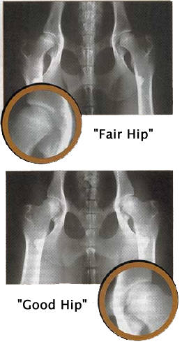
In the hip-extended view, the dog is laid on its back and the hind legs drawn backward, parallel to one and another (bottom right). This is the required view for submission to the Orthopedic Foundation for Animals (OFA).
Radiologists working for OFA evaluate the radiographs for two criteria. Movement of the head of the femur out of the acetabulum (in simple terms, the ball out of the socket) suggest joint laxity. Secondly, they look for arthritic change. The x-rays are scored Excellent, Good, Fair, Borderline, Mild HD, Moderate HD or Severe HD.
The latin definition of hip dysplasia is simply "faulty development of the hip." A Better definition in the 1960s, was, "a varying degree of laxity of the hip joint permitting subluxation during early life, giving rise to varying degrees of shallow acetabulum and flattening of the femoral head, finally inevitably leading to osteoarthritis."
The OFA assessment for HD has been hailed for many years as the best HD-rating system. Recently this status has been questioned. To assess the OFA method of scoring, Drs. Smith and McKelvie at the University of Pennsylvania sent radiographs to three radiologists - all board-certified - including one who reads radiographs for OFA itself, and then submitted these films to OFA. They went further by later sending the same x-rays to the same radiologists, a second time. The authors concluded that OFA testing techniques has "problems associated with wide variation in interpretation among radiologists."
Because of the concerns with the OFA technique, the University of Pennsylvania Hip Improvement Program (PennHIP) was developed. Though is is a radiographic technique, it is radically different from the standard OFA approach.
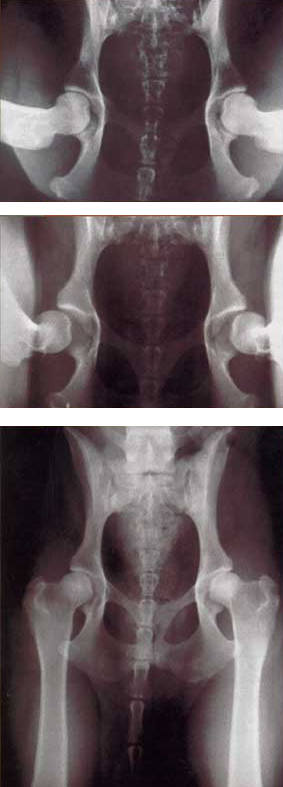
PennHIP positioning requires that the patient be anesthetized or very heavily sedated. Three radiographs are take; a compression view, where the hip is in a natural stance; a distraction view, where a custom-designed device if used to stress the hip joint to its natural limit; and a standard view, hip-extended view. Only veterinarians trained in the technique can take PennHIP radiographs and interpret them correctly.
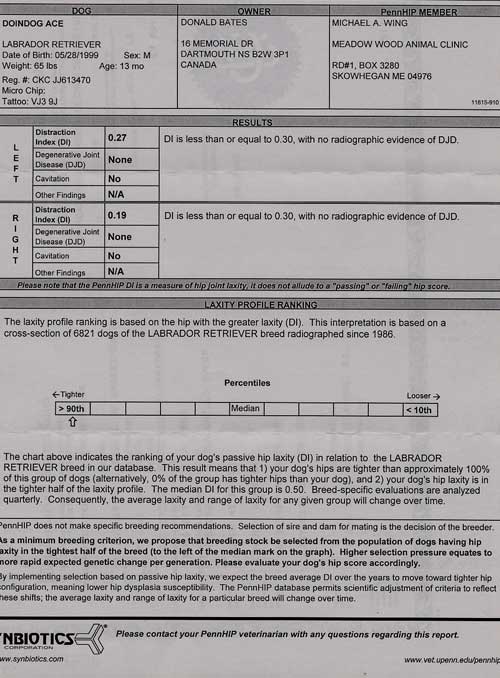
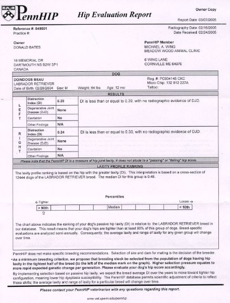
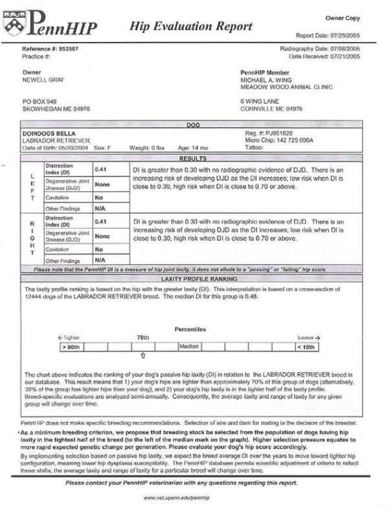
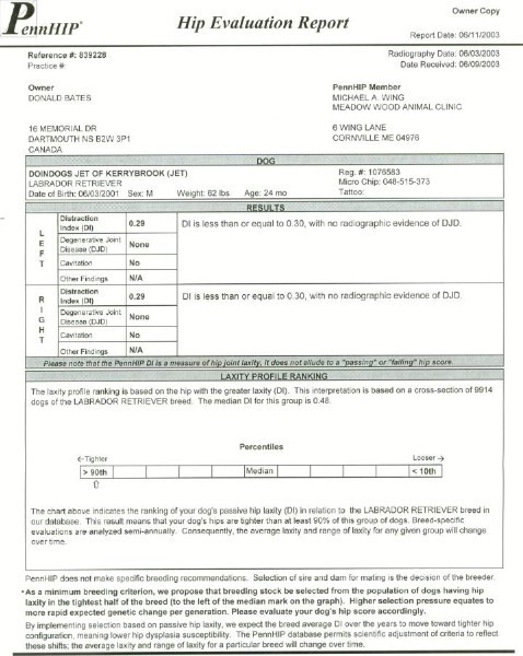
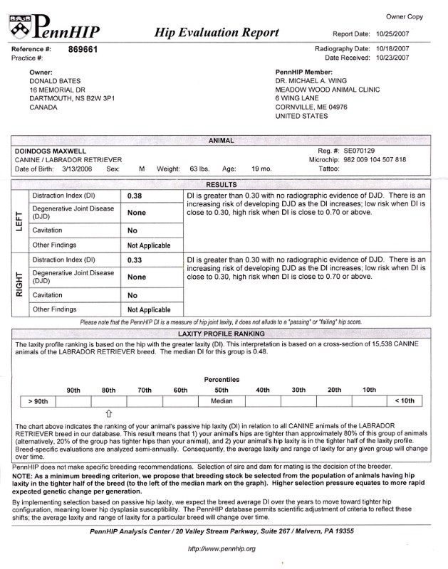
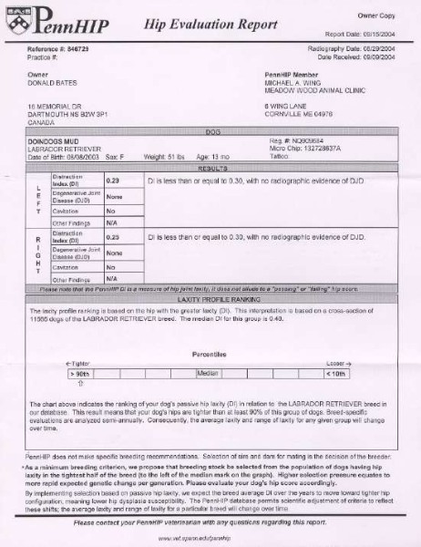
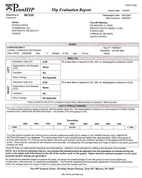
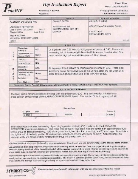
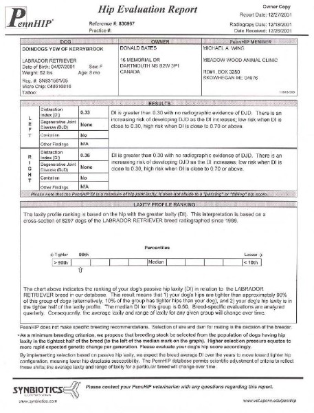
Once PennHIP radiographs are analyzed, the dog is assigned a distraction index. The lower rating, the less likely that the dog has HD and the lower his chance of developing osteoarthritis. Distraction indexes are not valid for comparing one breed to another but are valuable in selecting the better candidate within a breed based on hip architecture.
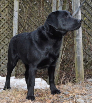
PennHIP reports for Doindogs Kennel Dogs:
Note: None of my dogs or any of their pups have developed Hip Dysplasia
How do the methods compare?
In one study, dogs that were rated OFA dysplastic (Mild, Moderate or Severe) also had distraction index greater than 0.3 (with a mean of 0.55). Both methods readily identified dogs with obvious hip-joint pathology; however, half the dogs 'cleared' by the OFA were considered by PennHIP to be susceptible to degenerative joint disease and at risk for developing arthritis. The PennHIP method identifies more dogs with a tendency for HD than the OFA assessment.
Because PennHIP reveals a higher incidence of HD, it is - according to the university that developed it - a tool that has a great potential to lower the frequency of HD if it is used as a selection criterion for breeding dogs.
OFA insists that its method reduces the incidence of HD and has a proven track record over the last two decades. The overall incidence of HD has been reduced.
Will PennHIP replace the current system of OFA certification? Even though it appears superior, there are a few certified veterinarians who can perform the procedure, so it will be slow to gain acceptance. As well, some breeders and veterinarians stand firmly behind the OFA procedure. A few breeders are hedging their bets by submitting their dogs to both forms of assessment.
In the end, it will be the dog-breeding community that will dictate which certification program is used in the future. They will choose the method that assists them in lowering the frequency of HD. If the PennHIP is ultimately desired, veterinarians will undertake the expense and Training needed to become PennHIP-certified.
Time will Tell
by Jeff Grognet, D.V.M., B.Sc.(Agr.) (August 2001)
Doindogs Note:
Please note that PennHIP is now an established method (2008)
I have become a big fan of the PennHIP method. One of the biggest advantages for me is that the dog can be certified as early as 20 weeks. I have had several dogs done by PennHIP and have found that the vets also prefer this method.
I have taken dogs to Dr. Michael WING, Skowhegan, ME, U.S.A. (207) 474-2938. Mike owns several labs and also believes the PennHIP to be a superior method.
Not many vets are able to perform PennHIP radiographs. Dr. Ernie PROWSE, an Orthopedic specialist is certified to perform PennHIP. Arrangments can be made though my vet Dr. RUSHTON at:
Westwood Hills Veterinary Hospital
4 Westwood Blvd Hubley Centre
Upper Tanallon, Nova Scotia
Canada, B3Z 1H3
Tel: 902-826-1933
Fax: 902-826-9466
home > articles > Assessing Hips (How the OFA and PennHIP methods compare)